 LoonaMed
Products/LoonaMed MRI
MRI, mapped
and comparable over time.
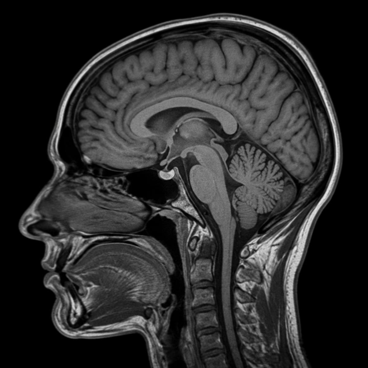
Load an MRI study and LoonaMed MRI maps the anatomy across sequences, measures each region and lines the results up with prior scans — you review, adjust and sign off.
- Input
- MRI series (DICOM)
- Output
- Region maps & volumes
- Follow-up
- Compared with prior studies
How it works
From series to follow-up.
SAG T1 · 176 slices · 1.0 mm
Series 1 of 4 · 0/176
Prior · Mar 2025
Volumes · cm³
| Total brain | — | 1,050–1,350 |
| Cerebellum | — | 120–160 |
| Brainstem | — | 24–33 |
| Ventricles | — | 10–40 |
Change since Mar 2025
| Total brain | — | 1,184 |
| Cerebellum | — | 142 |
| Brainstem | — | 28.4 |
| Ventricles | — | 23.8 |
MRI report
Study 2291
Generated automatically · Pending clinician review
Mapping
Major regions mapped across 176 slices
Volumetrics
Regional volumes within reference ranges
Follow-up
Ventricular volume +1.1% since Mar 2025
What's inside
Everything LoonaMed MRI does,
in one focused tool.
Multi-sequence studies
T1, T2, FLAIR and diffusion series are read together from one study, so you can move between them at the same position.
Precise anatomical mapping
Major brain regions are outlined across every slice and shown as clear overlays you can toggle, inspect and correct.
Regional volumetrics
Volumes are computed for each mapped region and shown alongside reference ranges, in consistent units.
Comparable over time
Add a prior study and LoonaMed MRI lines the two up, showing what changed and by how much since the last scan.
Structured reports
Mapping, volumes and changes are drafted into a structured report, ready to review, edit and export as PDF.
Clinician stays in control
Adjust any outline and the volumes and comparisons update instantly — nothing is final until you sign off.
Contact
Bring your next MRI.
Send us a few anonymised MRI studies — ideally with a prior — and we'll show you LoonaMed MRI working on your own cases.